

Private consultation

DR JASON EMER

SCROTAL
CALCINOSIS

ADVANCED EXPERTISE IN INTIMATE SKIN

Dr Jason Emer evaluates intimate skin nodules with a focus on diagnosis, procedural options and healing

Firm nodules in the scrotal skin deserve a clear diagnosis. Scrotal calcinosis involves calcium deposits in the skin, but not every lump in this area is calcinosis. An examination helps establish the cause and whether a procedure is appropriate.

SCROTAL CALCINOSIS / INTIMATE DERMATOLOGY

A PRIVATE CONCERN
A CLEAR WAY FORWARD

You may have lived with firm nodules for years before asking about them. The first step is a clear diagnosis and an explanation of what removal would mean for your skin.

Dr Emer assesses the number, size, depth and distribution of the nodules before planning treatment. Surgical excision, selected CO2 laser treatment or a combination may be considered. Your plan accounts for removal, the surrounding skin and healing.

Explore treatment and results

THE REASON BEHIND THE PLAN

WHAT SHAPES
THE REMOVAL PLAN?

The extent of the finding changes both the technique and the recovery discussion.

Confirm what the nodules are

Calcinosis involves calcium deposits in the skin. Other lumps can look similar, so the examination comes before a removal plan. The clinician decides whether further investigation is needed.

A photograph alone cannot establish the diagnosis.

Understand the condition
Match treatment to the distribution

Surgical excision removes affected tissue. CO2 laser treatment may be considered for selected nodules. Their size, depth and distribution help determine the approach, whether a combination is appropriate and whether treatment should be staged.

The plan may be staged when the extent of treatment calls for it.

Watch Dr Emer explain removal

Plan for the skin after removal

Wound care, swelling, activity and follow-up depend on the extent and method. Ask how scars and possible new nodules will be reviewed over time.

Recovery is individual. Obtain specific guidance about work, exercise and sexual activity.

Explore the care sequence

SCROTAL CALCINOSIS, EXPLAINED

UNDERSTAND
WHAT THE NODULE IS

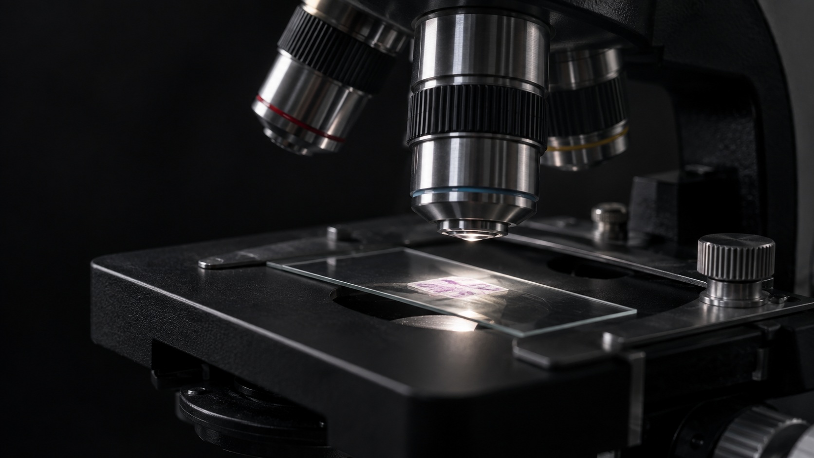
An assessment starts with the skin findings and your history. When tissue is removed, pathology can help identify the material and clarify the diagnosis.

Microscope and tissue slide illustrating pathological assessment

Start with
an examination

Not every firm skin bump is calcinosis. Size, location, symptoms and how the nodules have changed help guide the assessment and whether further evaluation is appropriate.

Tissue can provide
a clearer answer

Calcinosis involves calcium deposits within skin. Pathology may confirm this when tissue is sampled or removed. The microscope scene illustrates evaluation, not a result from a particular patient.

Match the plan
to the findings

The number and distribution of nodules, discomfort and skin available for closure affect the discussion. Ask about scars, wound care and the possibility of recurrence before choosing treatment.

FROM THE PRACTICE

SEE THE WORK
UNDERSTAND THE PLAN

Watch Dr Emer explain scrotal-calcinosis removal. The video includes intimate anatomy and procedure footage.

Scrotal calcinosis removal explained

Dr Emer discusses the condition and demonstrates treatment. Published April 10, 2026.

Open on YouTube

YOUR CARE, IN SEQUENCE

REMOVAL
THEN RECOVERY

A clear plan covers the procedure and the days that follow, including wound care, activity and the return visit.

Discuss your individual plan

Share the history

Describe the duration, growth, discomfort or discharge and any previous procedures. Tell the clinician about other health conditions and medications.

Understand the diagnosis

Ask what supports the diagnosis and whether further assessment or tissue examination is needed. A testicular lump needs a different evaluation.

Prepare for healing

Review the proposed technique, wound care, clothing, hygiene and restrictions on sex or exercise. Get instructions tailored to the extent of treatment.

Attend follow-up

Discuss how healing will be reviewed and what symptoms should prompt earlier contact. New nodules or recurrent concerns deserve reassessment.

THE QUESTIONS WORTH ASKING

LET’S GET
SPECIFIC

Open a question for the details. Bring your own questions to the consultation, too.

Is every scrotal lump calcinosis?

No. A clinician needs to establish where the lump is and what is causing it. Skin nodules and testicular lumps are different concerns.

What is scrotal calcinosis?

It involves calcium deposits forming nodules within the scrotal skin. Diagnosis should be confirmed clinically, with further assessment when appropriate.

Does it always mean high blood calcium?

Not necessarily. The clinician should decide whether your history or examination calls for additional testing.

Can the nodules be removed?

Procedural removal may be an option. Technique and staging depend on the number, size, location and skin involved.

Can it come back?

Recurrence is possible. Follow-up and realistic expectations should be part of the discussion.

Will there be a scar?

Scarring is possible with removal. Ask how the proposed technique balances treatment of the nodules with healing and the surrounding skin.

The Evolution of You™

YOUR NEXT STEP
CAN BE A CONVERSATION

Share how long the nodules have been present, whether they cause symptoms and what you would like to understand about removal.

Request your consultation

More about your treatment

DermNet: calcinosis cutis

Illustrations explain anatomy and treatment concepts. They do not show a patient’s diagnosis or treatment result.