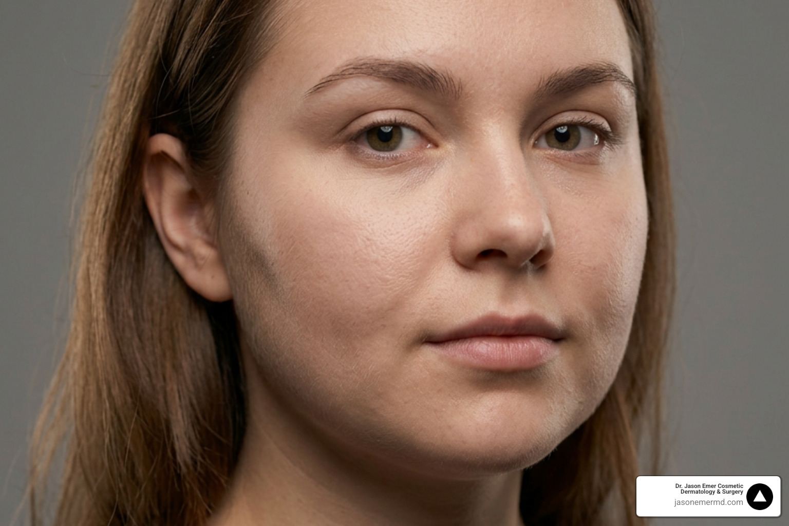
Buccal Fat Pad Removal: A Careful Approach to Cheek Contouring
A slimmer face is not always the right goal. Buccal fat pad removal can reduce deep lower-cheek fullness and accentuate cheekbone contours, but it requires careful evaluation of bone structure, tissue volume, and long-term aging patterns.
The buccal fat pad sits deep in the cheek between the cheekbone and jawline. During the procedure, a clinician reaches the pocket through a small incision inside the mouth to remove a conservative amount of fat. There is no external facial incision. Removing too much tissue can cause hollow or uneven contours as the face naturally loses volume over time.
Clinical evaluation examines:
- Origin of cheek fullness (buccal fat versus general weight or laxity)
- Cheekbone structure and midface support
- Existing facial narrowness, hollowness, or asymmetry
- Age-related volume shifts and long-term structural balance
- Surgical risks, including swelling, trismus, bleeding, and nerve injury
Jason Emer, MD FAAD approaches cheek reduction as part of a comprehensive facial-balance assessment. The decision to reduce cheek volume is determined during a consultation, where facial anatomy and skin quality are evaluated.
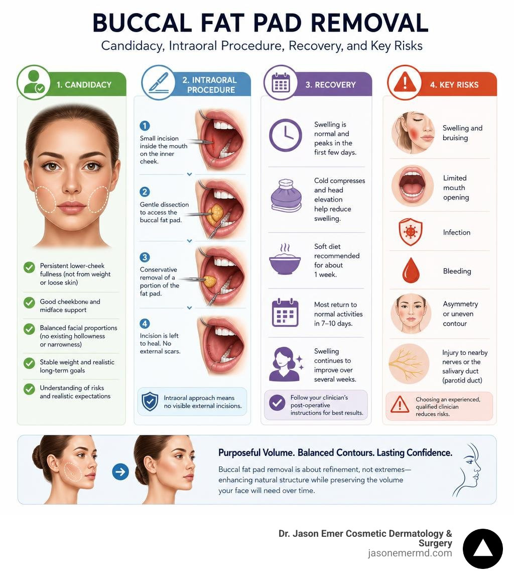
Buccal fat pad removal refines facial contours by addressing genetically persistent cheek volume
Deep facial fullness often resists traditional diet and exercise. For many patients, the lower cheeks remain full regardless of body weight, because the tissue responsible is not the same fat that responds to caloric change. The buccal fat pad, or Bichat's fat pad, is an encapsulated adipose structure in the lower midface. It consists of anterior, intermediate, and posterior lobes. The posterior lobe features four extensions, including the buccal extension responsible for anterior cheek fullness. Anatomical data show that the buccal extension and main body comprise roughly 50 percent of total pad volume.

Anatomical differences guide operative planning. A customized approach for female patients often emphasizes cheekbone projection and subtle submalar shading. Conversely, tailored contouring for male patients focuses on a defined, angular jawline. Volumetric variation ranges from 7.8 to 11.2 milliliters in males and 7.2 to 10.8 milliliters in females, with an average thickness of 6 millimeters. These numbers matter because they set a ceiling on how much tissue can reasonably be removed without disrupting the structural scaffolding of the cheek.
Anatomical considerations during buccal fat pad removal
The surgical space sits between the buccinator and masseter muscles. Clinical precision is required because the buccal branch of the facial nerve and the parotid duct run adjacent to the fat pad. Standard protocol relies on conservative extraction, typically removing only 3 to 5 cc of fat. Applying finesse techniques in fat excision preserves structural support and prevents excessive hollowing. Working within this narrow anatomical corridor is one reason the procedure is approached deliberately rather than aggressively; removing slightly too much tissue is far harder to correct than removing too little, since the fat pad does not regenerate once excised.
Surgical technique and procedural safety minimize complication rates and protect delicate structures
A careful technique limits tissue trauma. The procedure is performed through a small intraoral incision inside the cheek mucosa, avoiding external scarring. Before entering the buccinator space, clinicians identify Stenson's duct opening to reduce injury risks. The fat is gently teased forward and trimmed without traction before closing with absorbable sutures. Because the incision sits inside the mouth, saliva and oral bacteria are ongoing considerations during the healing process, which is part of why oral hygiene instructions are emphasized during recovery.
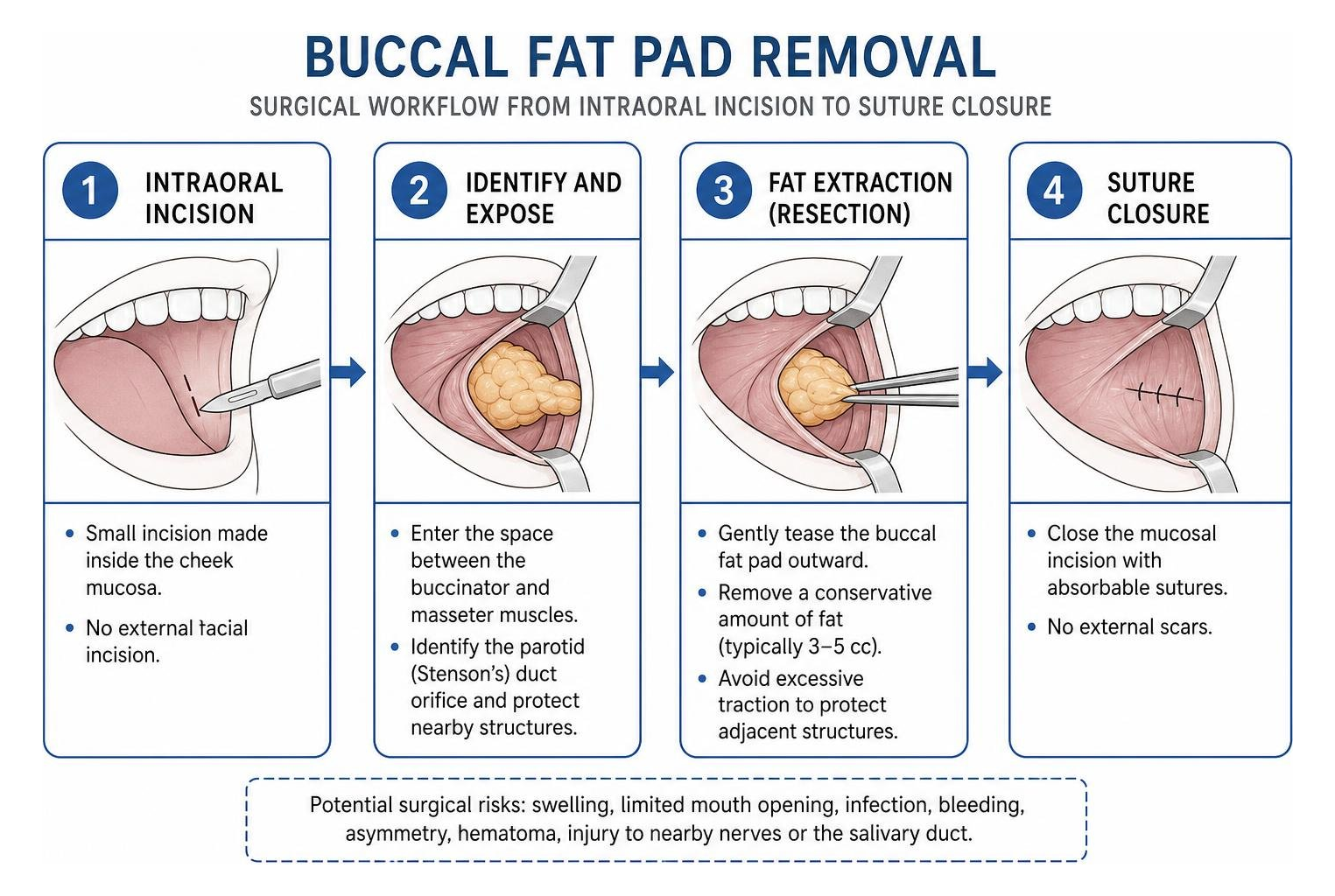
Reported complication rates range from 8.45% to 18%. Potential risks include hematoma, bleeding, salivary retention from duct injury, motor changes from nerve trauma, infection, asymmetry, swelling, and trismus. These figures are a real limitation of the procedure, and they are part of why a conservative extraction volume is preferred over an aggressive one. A post-operative clinical recovery evaluation confirms that aftercare is essential to risk management.
Postoperative recovery and tissue healing timelines
Healing occurs in phases, and recovery should be treated as part of the treatment plan rather than an afterthought. Swelling peaks during the first four days and improves substantially by day 15. Temporary stiffness or limited mouth opening can occur if the buccinator muscle is irritated. According to clinical overviews of recovery and patient care, jaw mobility generally returns to baseline by day 30. Postoperative care includes cold compresses for 24 to 48 hours, soft foods, and antiseptic oral rinses. Patients are generally advised to avoid strenuous exercise and excessive jaw movement, such as wide yawning or chewing tough foods, during the initial healing window. Following these steps closely tends to correlate with a smoother recovery, though timelines vary based on individual tissue response and the extent of tissue removed during surgery.
Strategic midface sculpting requires evaluating structural aging to avoid excessive volume loss
Cheek contouring must account for aging. The buccal fat pad expands into early adulthood and gradually declines over subsequent decades. If a face exhibits narrow cheeks, limited cheekbone projection, or early hollowness, reducing volume can lead to an aged or gaunt appearance. This is a genuine tradeoff of the procedure: buccal fat pad removal is not reversible, and tissue that is taken out cannot be replaced without a separate volumizing treatment later.
Clinical decisions may involve conservative reduction, delaying surgery, or choosing non-surgical alternatives, including the option of doing less and monitoring the face over time rather than intervening immediately. When contouring is appropriate, Dr. Emer may evaluate specialized buccal fat pad removal, broader facial sculpting, or facial fat grafting to restore support. Each of these paths carries a different risk and recovery profile, and the choice between them is made only after a full facial assessment.
When cheek reduction is combined with lasers or energy devices, skin type is critical. Darker skin tones face higher risks of post-inflammatory hyperpigmentation, requiring conservative device settings and specialized treatment sequencing. This is one reason combination treatment plans are staged carefully rather than performed all at once, since surgical healing and energy-based treatments both affect the same skin and tissue.
Long-term aging dynamics following buccal fat pad removal
Complete excision removes structural support that is difficult to restore. Preserving the main body of the pad while reducing only the protruding buccal extension helps maintain balanced submalar contours over time. Because facial fat naturally redistributes with age, a face that looks well-balanced immediately after surgery may look different a decade later, which is why long-term structural planning is discussed as part of the evaluation rather than judged only on immediate results.
Conclusion
Facial refinement requires a careful balance between soft tissue reduction and structural preservation. Determining candidacy, surgical staging, and procedural volume relies on comprehensive anatomical evaluation during a consultation for comprehensive facial surgery.
What to remember
- Buccal fat reduction requires conservative extraction of 3 to 5 cc to prevent midface hollowing.
- Complication risks range from 8.45% to 18% and include localized edema, trismus, and potential duct or nerve injury.
- Long-term facial aging naturally reduces buccal pad volume, making cheekbone support a vital structural consideration.
This content is for general education and does not provide a diagnosis, determine candidacy, or replace a consultation with a licensed clinician.
References
Barron Davis, Marc Serra. "Buccal Fat Pad Reduction." StatPearls Publishing, 2022. NBK576413.
This resource provides general educational information. Treatment recommendations depend on your health, goals, and consultation with a qualified clinician.