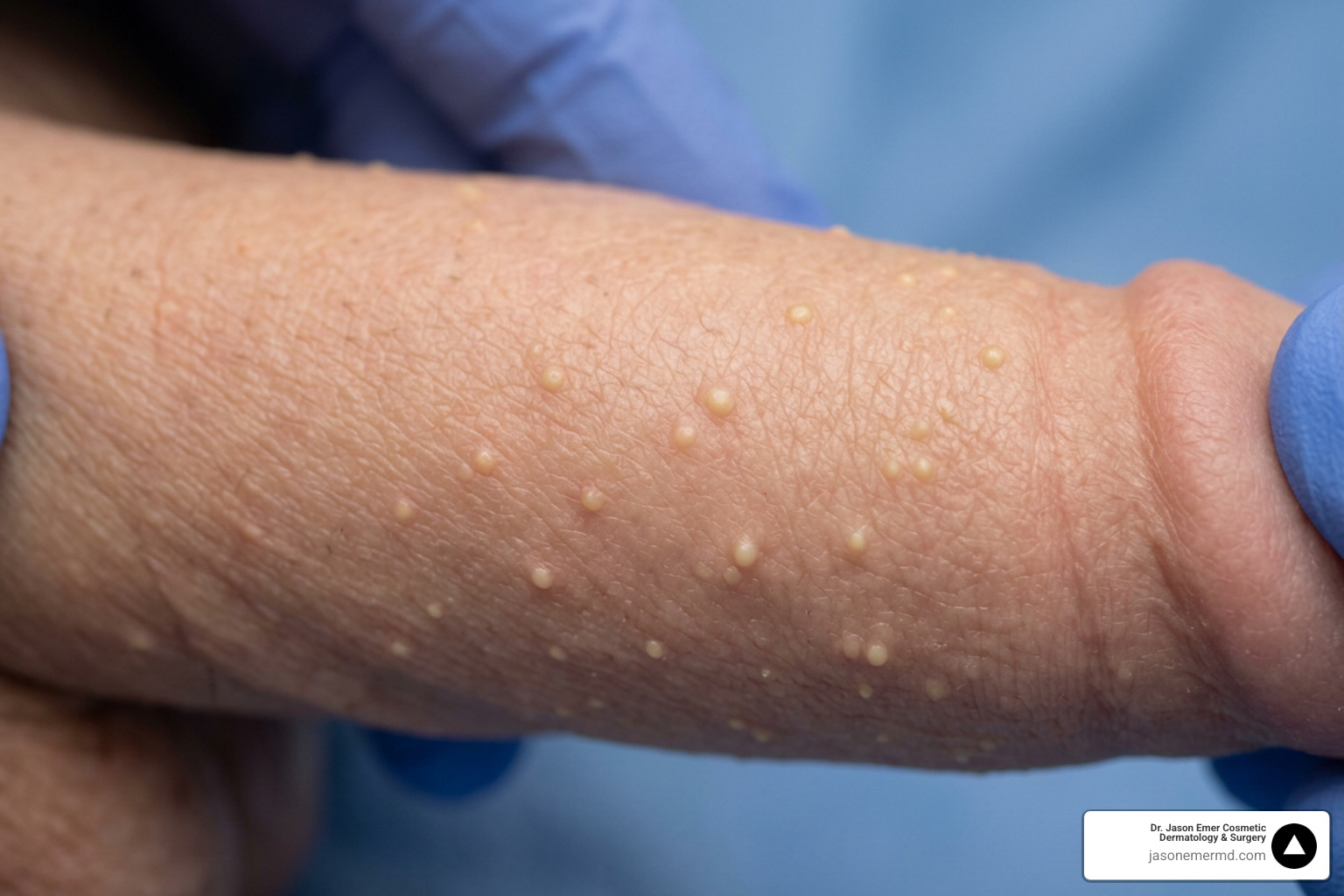
Penile Fordyce spots are usually benign, not an STI
Most people assume any new bump on the penis signals an active sexually transmitted infection. The better clinical model starts elsewhere: look at the underlying tissue first. Penile Fordyce spots are visible oil glands, not an infection, and identifying them takes three simple steps. First, examine the pattern: they appear as small, smooth white or yellowish papules clustered on the shaft, prepuce, or scrotum, becoming more visible when skin is stretched. Second, check the texture: unlike genital warts, they remain smooth rather than rough, and unlike herpes, they never blister or scab. Third, monitor them: they require no treatment unless they change, become painful, or need diagnostic confirmation during an in-person exam.
These ectopic sebaceous glands represent a normal anatomical variant. They reside within the dermis without hair follicles, allowing lipid contents to show through the thin penile skin. Nearly every adult carries some version of this tissue somewhere on the body, whether on the lips, cheeks, or genital skin, and the penile presentation is simply one of the more noticeable locations because of how thin and mobile that skin is.
Anxiety about new genital bumps is common, and understandably so. Sexually transmitted infections are a real concern, and self-diagnosis from a mirror rarely settles the question with confidence. That is why a structured look, rather than a snap judgment, matters. Pattern, texture, and behavior over time each carry more diagnostic weight than the mere presence of a bump.
Three practical habits protect the skin barrier:
- Never squeeze or pop the bumps. Manual trauma risks secondary bacterial infection, swelling, and scarring.
- Use gentle hygiene. Wash with mild cleansers and avoid harsh astringents or tight synthetic friction.
- Seek clinical evaluation if lesions itch, bleed, or change.
Dr. Emer evaluates penile Fordyce spots carefully, ensuring patients understand their benign anatomy before considering conservative care or procedural options. That evaluation typically involves a brief visual exam, a short discussion of history and any changes noticed, and, when needed, magnified inspection to confirm the diagnosis without guesswork.
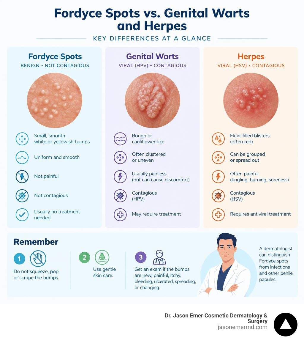
Penile Fordyce spots are benign anatomical variations that can resemble infections
Penile Fordyce spots represent heterotopic sebaceous glands without associated hair follicles. Present from birth, these structures become prominent during puberty under androgenic stimulation and are non-contagious. They do not spread through sexual contact, and they do not indicate poor hygiene or an underlying illness, a point worth repeating because so much of the anxiety around these bumps comes from an assumption of contagion that simply does not apply here.
A clinical assessment by a dermatologist distinguishes these glands from viral infections, as documented in educational resources like Fordyce spots: Quick and Informative guidance — DermNet. Lesions present as 1 to 3 millimeter, non-tender, creamy-yellow or whitish papules clustered across the penile shaft, prepuce, or scrotum. Unlike human papillomavirus (HPV) lesions, which display rough surfaces, Fordyce spots remain smooth and uniform without blistering or crusting.
Visual inspection remains standard. Diagnostic confidence improves with ultraviolet-induced fluorescence dermatoscopy, where sebaceous clods emit distinct blue-green fluorescence, as explored in a study on differentiating Fordyce spots from their common simulators using ultraviolet-induced fluorescence dermatoscopy. Histologic punch biopsy is rarely required, and is generally reserved for cases where the presentation is atypical or where a patient has a personal history that raises the index of suspicion for something else entirely.
Because these glands are a normal feature of skin anatomy rather than a disease process, their number and visibility can vary widely from one person to another. Some men have only a handful of scattered spots; others have dense clusters covering much of the shaft. Neither pattern is abnormal, and neither pattern predicts any future health issue. The variation is simply a reflection of how much sebaceous tissue was distributed to that area during development.
A clinical evaluation distinguishes penile Fordyce spots from other genital papules
Morphology and anatomical distribution guide the differential diagnosis of benign genital lesions. Because multiple skin structures occur in male pelvic anatomy, an expert physical examination confirms whether bumps represent sebaceous glands or alternative proliferations.
Pearly penile papules represent one of the most frequent physiological findings. These lesions appear as small, pearlescent, dome-shaped angiofibromas aligned along the corona of the glans penis and the coronal sulcus. While pearly penile papules remain restricted to the coronal margin, Fordyce spots occupy the mid-to-proximal shaft, prepuce, and scrotum. Tyson glands present specifically at the coronal sulcus flanking the frenulum.

Vascular and follicular conditions must also be considered. Angiokeratomas of Fordyce manifest as dark red or violaceous vascular ectasias that may bleed after minor friction. Sclerosing lymphoceles present as firm, cord-like swellings in the coronal sulcus following vigorous activity. Epidermal inclusion cysts and folliculitis display inflammatory margins or central puncta, whereas ectopic sebaceous glands remain indolent.
Accurate evaluation prevents unnecessary anxiety and protects patients from inappropriate over-the-counter wart removers or caustic acids that cause chemical burns on delicate penile tissue.
Conservative care and professional procedures can address cosmetic concerns about Fordyce spots
Because Fordyce spots represent normal physiological structures, doing nothing remains the primary medical recommendation. They cause no physiological harm and do not impair sexual function. For individuals experiencing aesthetic distress, clinical management focuses on education, barrier support, or controlled procedural modalities.
In-office procedural options target visible glandular structures with high precision. Modalities include ablative carbon dioxide ($CO_2$) lasers, erbium:YAG ablation, targeted electrodessication, radiofrequency, and micro-punch excision. When utilizing energy-based devices on genital skin, skin phototype requires strict evaluation. Darker skin tones carry an elevated risk of post-inflammatory hyperpigmentation (PIH), hypopigmentation, and scarring due to heightened melanocytic sensitivity to thermal injury. For higher Fitzpatrick skin types, conservative energy settings, cautious test spots, extended treatment intervals, and customized staging minimize thermal injury.

Recovery requires strict post-procedure wound management. Re-epithelialization spans 5 to 21 days depending on ablation depth and healing rates. Mild localized swelling, light crusting, and transient erythema are anticipated procedural trade-offs. Patients must maintain clean wound margins, apply soothing occlusive ointments, and avoid sexual activity during recovery to prevent mechanical shear stress.
Non-procedural alternatives include topical retinoids, though they can provoke local irritant contact dermatitis on thin genital skin. Patients seeking aesthetic management can consider an individualized clinical consultation for pearly penile papules and Fordyce spots to determine procedural suitability.
What to remember
- Penile Fordyce spots are benign, non-contagious ectopic sebaceous glands present in the vast majority of adults.
- Professional examination with dermatoscopy easily differentiates Fordyce spots from sexually transmitted infections and other benign papules.
- Procedural removal via laser or electrodessication is strictly cosmetic, carries risks such as post-inflammatory pigment alteration, and requires personalized clinical evaluation.
This content is for general education and does not provide a diagnosis, determine candidacy, or replace a consultation with a licensed clinician.
This resource provides general educational information. Treatment recommendations depend on your health, goals, and consultation with a qualified clinician.